Actin and Myosin
October 6, 2011 in Protein
Actin and Myosin
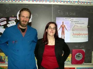
On Monday, February 18th, 2008, the students in Mrs Shuster’s third grade class at École F.A.C.E. School flexed their muscles, as university students Marie-Eve Chartrand (BFA Art Education, Concordia U.) and Stephan Brodeur (B.Sc., U. Montréal) presented the proteins of movement “actin and myosin” in our fourth Molecules of Life Project (MLP) in Montreal.
Stephan and Marie-Eve were quick to put the students to work and asked them first to raise their chairs, close their eyes and imagine that they were holding an elephant sitting on each of their chairs.
This simple experiment led to a profound discussion on how the brain controls the muscles of the body and how energy is needed for the muscles to work.

Switching to the cellular level, Marie-Eve constructed a model of the cell using a zip-lock plastic bag filled with water, a marble for a nucleus and string, to describe the micro-filaments that the cell uses in order to move.

Stephan showed an experiment in which a chemical reaction created gas that popped the top off a bottle in order to iterate the point that chemical energy can be used to create physical motion.
Employing different kinds of beaded strings, Stephan and Marie-Eve described the protein actin and how it forms similarly a string of protein beads on which the protein myosin climbs. Using styrofoam balls and pipe-cleaners, the students constructed their own model of actin fiber.

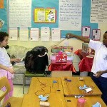
The movement of myosin along actin was then presented by the construction of a micro-filament model from a paper plate and string. The students first cut out from the plate an “X”-like shape that represented the protein myosin, and punched holes at the four arms of their myosin model.

Setting in the actin fibers on which their myosin would move, the students threaded cord through the four holes. Mimicking the movement of the proteins in muscle fibers when the brain signals them to move, the students used their own energy and pulled the actin cords, to propel their myosin to climb up the actin. In the same way that muscles return to the relaxed state when there is no longer signal to contract, when the students energy stopped pulling, the myosin slid back down along the actin fiber to a resting position.

Playing with their actin and myosin models, the students grasped the importance of energy for the movement of the two proteins necessary for muscle contractions and motion.

Marie-Eve and Stephan explained how muscle motion can be stopped by diseases that effect the actin and myosin filaments such as muscular dystrophy as well as when signal from the brain is blocked due to spinal cord injury . Finally, we discussed how muscles can grow weak without regular exercise and how a diet containing foods rich in protein was essential for building new actin and myosin filaments. Using their muscles to give a round of applause, the students thanked team actin and myosin for a moving MLP experience.
For an animation on actin and myosin, micotubules, cellular movement and muscle movement see:
www.wiley.com/college/pratt/0471393878/student/animations/actin_myosin/actin_myosin.swf
For a cartoon on the movement of actin and myosin see:
www.accessexcellence.org/RC/VL/GG/ecb/myosin_and_actin_model.html
For a descriptive animation of how muscle movement occurs see: