Ink Composition and Chromatography

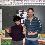
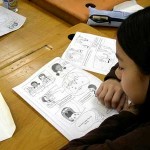
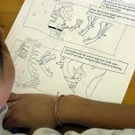
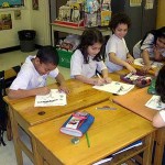
Molecules of Life and l’École de Manga Japonais de Montreal (The Montreal Manga School) team

An Elementary School Introduction to Carbon (part 2)- The Video

The following video is intended primarily as an aid for elementary school teachers and parents to fa

An Elementary School Introduction to Carbon (part 1)- The Video

The following video is intended primarily as an aid for elementary school teachers and parents to fa


Antibodies

October 6, 2011 in Protein

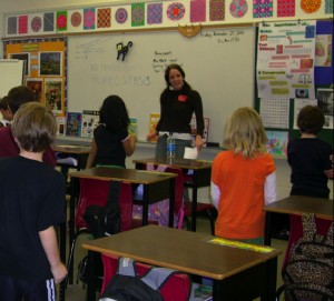
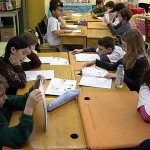
On Monday, February 11th, 2008, the students in Mrs Shuster’s third grade class at École F.A.C.E. School were infected by the presentation by university students Jesse Trubiano (BFA Art Education, Concordia U.) and David Sabatino (PhD. McGill ’07, PDF, U. Montréal), as they learned about antibodies the “Protein of Life” in our third Molecules of Life Project (MLP) in Montreal.

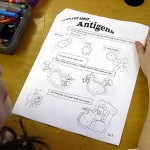
Jesse and David introduced us first to antigens (i.e. germs, bacteria and pollen) as they presented the immune system and explained the importance of washing our hands before eating.
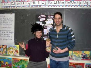
To demonstrate how quickly germs spread in the absence of hygiene, David guided two volunteers in experiments using sets of agar plates (Petri dishes containing the essential nutrients required to culture bacteria). One student washed carefully her hands before touching the plate labeled “clean”. The other student shook hands with 5 other students before touching the plate labeled “infected”. The plates were covered and stored in a dark warm place for the class to examine over the span of a week, to see what happens.
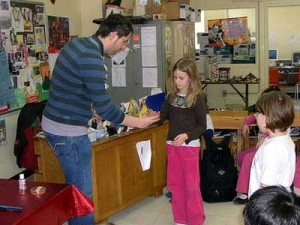
 

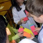
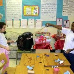
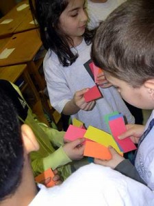
To illustrate the spread of germs and the importance of immunity to help prevent infection, the students were next given envelopes containing colored cards and asked to trade cards with one another for 2 minutes. Although most students were instructed that they could not refuse a trade, those students having a white “immunity” card were told that they could refuse exchanges of the white card. Once the time had elapsed, the children returned to their desks, examined their cards and those with a red card were asked to go to the back of the room, because they were infected with a cold germ. Those who also possessed a white card were told they could sit back down because they had immunity. The rest were asked to act out the symptoms of a cold until given purple “medicine” cards to cure their illness.


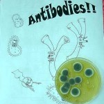
Jesse and David then distributed the “Immune System Comic Book featuring Antibodies” and the students were guided through the immune system as they took turns around the classroom reading about antigens, B-cells, antibodies and white blood cells, as well as the importance of vaccinations and hygiene.
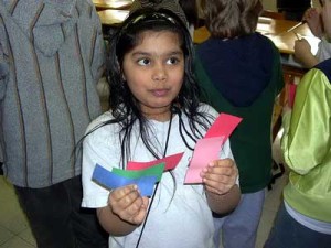
Antibodies were presented as the guard dogs of the immune system.
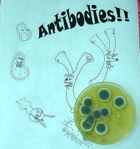
Finally, as the students colored in the comic book, we discussed the Y-shape of antibodies and how the arms of the Y serve to specifically recognize different germs.


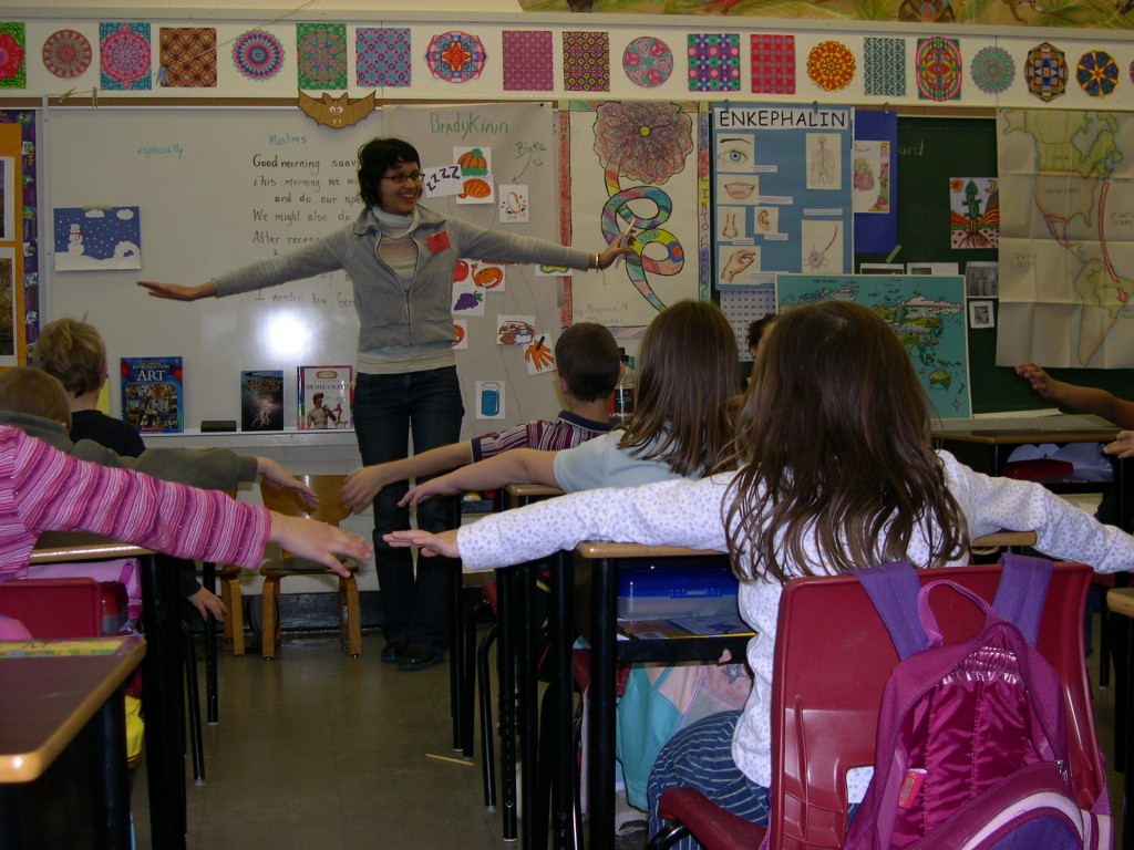
Mimicking antibodies all raised their arms in a Y shape to thank team antibody for an important MLP lesson on what it takes to stay safe from infectious germs.


For reviews of the immune system see: www.emc.maricopa.edu/faculty/farabee/biobk/BioBookIMMUN.html

and

http://uhaweb.hartford.edu/BUGL/immune.htm

For an animation on how the immune system works: www.doereport.com/generateexhibit.php?ID=15529&ExhibitKeywordsRaw=&TL=1&A=
 

Albumin

October 6, 2011 in Protein

On Thursday, March 20th, 2008, the students in Mrs Shuster’s third grade class at École F.A.C.E. School were stuck on “albumin” as university students Christina Thomson (MFA Art Education, Concordia U.) and William Bechara (B.Sc., U. Montréal) presented our sixth Molecules of Life Project (MLP) in Montreal.

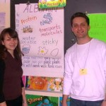
Christina and William discussed how like the school bus, albumin can serve as a molecule bus for transporting nutrients, metals, fats and other compounds by way of the blood through the body. Showing a poster to describe the structure and function of albumin, they mentioned how albumin is sticky and can serve like a sponge to absorb different molecules.

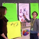
As a prelude to an experiment to discuss “solubility”, the students were asked to place “X” marks or check marks on another poster which reviewed laboratory safety practices.

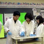
In proper laboratory safety attire, the students performed an experiment to model how albumin can help bring molecules into water and make them “soluble”. First iodine was added to water and remained “insoluble”, sitting like a rock in the bottom of the flask. Then a white powder (the amino acid cysteine) was added to the flask, and within seconds the iodine and powder both went into the water solution which became clear.

Asking students to attach pictures to another set of posters, Christina and William reviewed albumin’s uses in medicine, cooking and art. For example, Doctor’s use albumin to make band-aids stick to skin and to help medicine go into solution. Cooks use the cohesive properties of egg white albumin to make angel-food cake and soufflés.


Moreover, painters such as Sandro Botticelli used egg albumin to make tempera paint pigments more water soluble and more sticky when painting pictures such as “The Birth of Venus” and “La Primavera”.


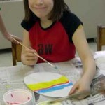
Inspired by Botticelli and the Easter Bunny, the students used tempera paints to color their own egg shape canvases.


Finally, we discussed the fact that allergies of different foods (i.e., eggs, peanuts, wheat, and milk) may be due to their different forms of albumin.

We thanked Christina and William for today’s MLP presentation which the students sponged-up like albumin.


For more information on albumin see:

http://en.wikipedia.org/wiki/Albumin

On the structure of albumin:

www.friedli.com/research/PhD/chapter5a.html

For details on the things that albumin binds to go to:

www.albumin.org/

For the Botticelli project see:

www.eggtempera.com/botticelli/botticelli.html

On albumin and food allergies see:

http://www.health.discovery.com/centers/allergyasthma/foodallergy/food.html

Actin and Myosin

October 6, 2011 in Protein

Actin and Myosin

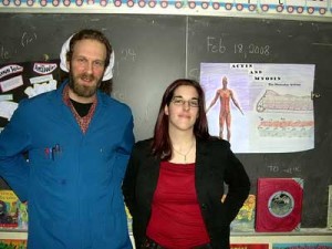
On Monday, February 18th, 2008, the students in Mrs Shuster’s third grade class at École F.A.C.E. School flexed their muscles, as university students Marie-Eve Chartrand (BFA Art Education, Concordia U.) and Stephan Brodeur (B.Sc., U. Montréal) presented the proteins of movement “actin and myosin” in our fourth Molecules of Life Project (MLP) in Montreal.

Stephan and Marie-Eve were quick to put the students to work and asked them first to raise their chairs, close their eyes and imagine that they were holding an elephant sitting on each of their chairs.

This simple experiment led to a profound discussion on how the brain controls the muscles of the body and how energy is needed for the muscles to work.


Switching to the cellular level, Marie-Eve constructed a model of the cell using a zip-lock plastic bag filled with water, a marble for a nucleus and string, to describe the micro-filaments that the cell uses in order to move.

Stephan showed an experiment in which a chemical reaction created gas that popped the top off a bottle in order to iterate the point that chemical energy can be used to create physical motion.

Employing different kinds of beaded strings, Stephan and Marie-Eve described the protein actin and how it forms similarly a string of protein beads on which the protein myosin climbs. Using styrofoam balls and pipe-cleaners, the students constructed their own model of actin fiber.


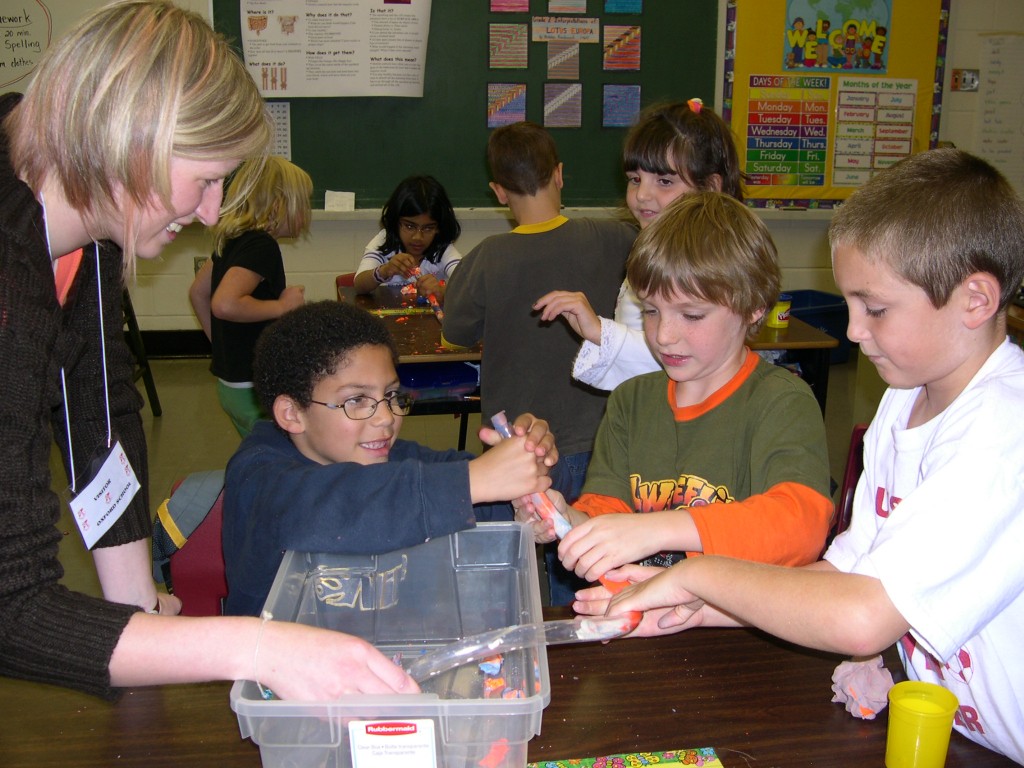
The movement of myosin along actin was then presented by the construction of a micro-filament model from a paper plate and string. The students first cut out from the plate an “X”-like shape that represented the protein myosin, and punched holes at the four arms of their myosin model.

Setting in the actin fibers on which their myosin would move, the students threaded cord through the four holes. Mimicking the movement of the proteins in muscle fibers when the brain signals them to move, the students used their own energy and pulled the actin cords, to propel their myosin to climb up the actin. In the same way that muscles return to the relaxed state when there is no longer signal to contract, when the students energy stopped pulling, the myosin slid back down along the actin fiber to a resting position.


Playing with their actin and myosin models, the students grasped the importance of energy for the movement of the two proteins necessary for muscle contractions and motion.


Marie-Eve and Stephan explained how muscle motion can be stopped by diseases that effect the actin and myosin filaments such as muscular dystrophy as well as when signal from the brain is blocked due to spinal cord injury . Finally, we discussed how muscles can grow weak without regular exercise and how a diet containing foods rich in protein was essential for building new actin and myosin filaments. Using their muscles to give a round of applause, the students thanked team actin and myosin for a moving MLP experience.


For an animation on actin and myosin, micotubules, cellular movement and muscle movement see:

www.wiley.com/college/pratt/0471393878/student/animations/actin_myosin/actin_myosin.swf


For a cartoon on the movement of actin and myosin see:

www.accessexcellence.org/RC/VL/GG/ecb/myosin_and_actin_model.html


For a descriptive animation of how muscle movement occurs see:

www.blackwellpublishing.com/matthews/myosin.html