Ion Channel Proteins

October 6, 2011 in Protein

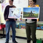
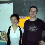
On Monday, February 25th, 2008, the students in Mrs Shuster’s third grade class at École F.A.C.E. School were charged-up by the discussion of university students Christine Sherrington (BFA Art Education, Concordia U.) and Nazim Ait Yahia (B.Sc., U. Montréal) as they presented “ion channel proteins” in our fifth Molecules of Life Project (MLP) in Montreal.

Christine and Nazim ask the students many questions and created a discussion that illuminated the presence of ions in food, such as calcium in milk, and sodium and chloride in salt, as well as the importance of ion channel proteins for controlling the flow of such ions into the cells of the body.

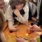
Employing a battery, two wires and a light bulb to create a circuit, Nazim illuminated the students with thoughts on the flow of ions from high to low charge and related ion channels to switches that regulate the flow of ions in and out of cells.

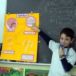
Showing an animation, Christine discussed how the ion channels form protein pores through the lipid (fatty) membrane of the cell.

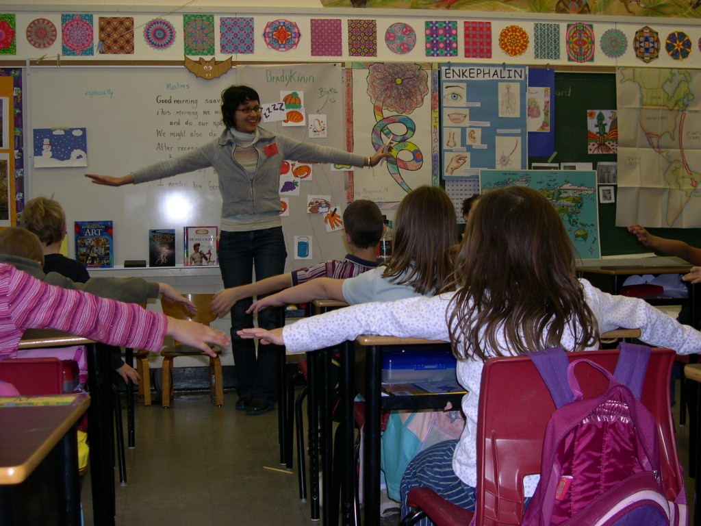
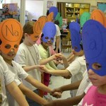
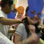
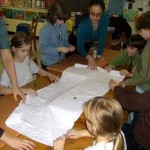
The students then tied on masks to become ions, and aligned themselves into two lines touching hands with their partners to form an ion channel protein.  Listening to Latin rhythms, the students danced the “ion channel salsa” in which they took turns being passed along the channel as ions, and passing their fellow students through the channel behaving like the side-chains of the amino acids which control the flow of ions in the ion channel protein.   Like ions flowing into a cell to form salts, the salsa dancers were dancing the mambo in the channel until the music stopped and the gates of the ion channel closed.  When the music resumed, the gates reopened and the dancers continued dancing into the cell.

…cha, cha, cha, mambo, mambo, mambo, salsa, salsa, salsa, ion, ion, ion, stop!

Finally, we discussed the importance of ion channels in regulating the functions of different cells and tissues, such as the contractions of muscle cells, the rhythm of the heart beat  and the flow of signals through the nervous system.  Experiencing the great potential of this learning experience, we thanked team ion channel for regulating a positive flow of MLP movement.


For some  Neuroscience For Kids:

http://faculty.washington.edu/chudler/ap.html


For a neat animation on ion channels involved in heart action:

http://www.cellsalive.com/channels.htm


for a basic review on ion channels:

http://www2.montana.edu/cftr/IonChannelPrimers/beginners2.htm


Hemoglobin

October 6, 2011 in Protein

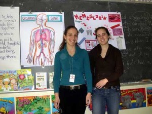
On Monday, February 4th, 2008, the students in Mrs Shuster’s third grade class at École F.A.C.E. School were magnetized by the presentation by university students Sabrina Beiba (BFA Art Education, Concordia U.) and Caroline Proulx (M.Sc., U. Montréal) as they learned about the importance of iron in hemoglobin the “Protein of Life” in our second Molecules of Life Project (MLP) in Montreal.

Sabrina and Caroline led a discussion that covered nutrition, the circulatory system and the transport of oxygen and carbon dioxide by iron in red blood cells.

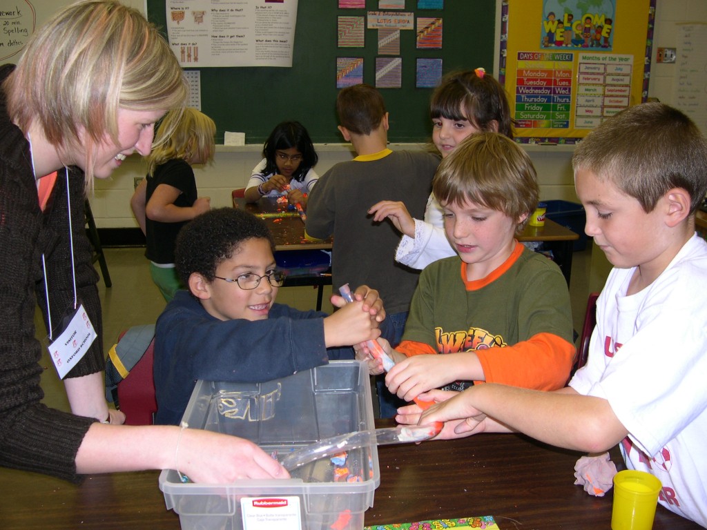
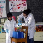
After refreshing the students on laboratory safety, Caroline helped a student volunteer perform the ferric chloride test in which a brown suspension of iron chloride in water was added to a suspension containing an amino acid (the building blocks of proteins) producing a bright yellow solution, which drew everyone’s attention.


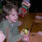
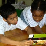
Packaging materials from various foods were distributed and the students were asked to read the Nutrition Facts and to determine which foods contained the most iron. Sabrina showed the students pictures of different foods and reminded the students that foods like raisins and spinach were good sources of iron.


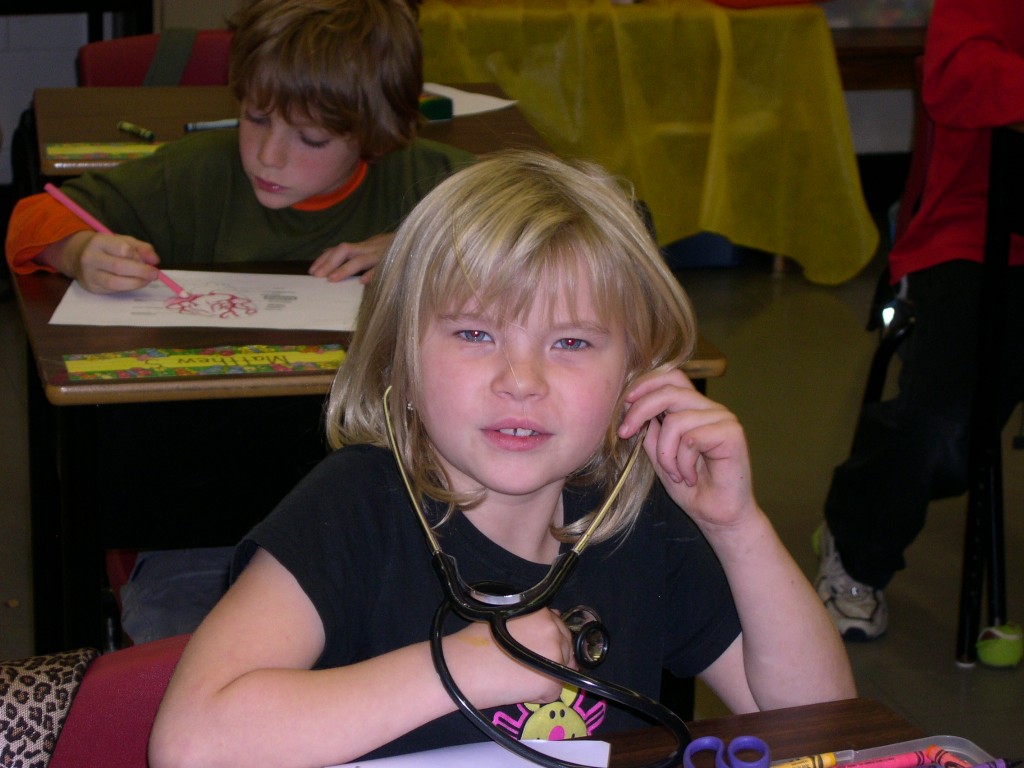
Sabrina and Caroline then led the students through the circulatory system. The students were first asked to each make a fist in order to perceive the size of their hearts, which pump blood throughout the body. Like tiny school buses which pick kids up and drive them to school and back, red blood cells were described as transportation machines which use iron contained in the protein hemoglobin to carry oxygen from the lungs to the cells and carbon dioxide from the cells back to the lungs. Like a fire requires oxygen to burn wood producing carbon dioxide, light and heat, the cells use oxygen to burn carbohydrates and sugars making carbon dioxide and energy.

Studying iron as an element which can grab onto gasses like oxygen and carbon dioxide as well as building blocks like amino acids, the students were instructed on how to use iron as a pigment for dyeing fabric. Employing clips and other metal objects made from iron, the students used rust (iron oxide) to bind to cotton as they worked in teams to prepare four banners.


They pleated the fabric. Clipped the pleats together using metal clips. In some cases, they wrapped iron objects with the fabric which was secured with clips.


Finally, they soaked the bound-up cotton fabrics in water. In one case, they added a little vinegar to see what might happen as well. The next day, they removed the clips and saw that the rust had dyed their banners an orange-brown color.


With some extra time, Caroline and Sabrina described how a modification of one amino acid in the protein hemoglobin can cause blood cells to change their shapes from disc-like cells into sickle shaped cells in the disease sickle cell anemia. By passing down a tube round chips to represent normal red blood cells and shell-shaped macaroni to represent sickle cells, they demonstrated how the sickle-shaped macaroni could block the flow through the tube, in the way that sickle cells may inhibit blood flow in sickle cell anemia, and explained that this could cause less oxygen to flow to the cells, which results in less energy.


WIth five more proteins scheduled this year, we thanked team hemoglobin for an iron-clad MLP performance at FACE.


For more information see:

Ferric Chloride Test: www.wellesley.edu/Chemistry/chem211lab/Orgo_Lab_Manual/Appendix/ClassificationTests/phenol_amine_nitro.html

The circulatory system: www.globalclassroom.org/hemo.html

Rust Dyeing: www.prairiefibers.com/Rust%20Dyeing.htm

Sickle Cell Anemia: www.kidshealth.com/teen/diseases_conditions/blood/sickle_cell_anemia.html


GPCRs

October 6, 2011 in Protein

Dear MLP enthusiasts,

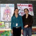
On Monday, April 7th, 2008, the students in Mrs Shuster’s third grade class at École F.A.C.E. School gained a better appreciation of their sense of smell as university students Catherine-Emmanuelle Drapeau (BFA Art Education, Concordia U.) and Tarek Kassem (Ph.D., U. Montpellier ’00) presented “G-protein coupled receptors” (GPCRs) in our seventh Molecules of Life Project (MLP) in Montreal.

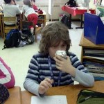
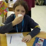
Catherine and Tarek distributed ten samples of boxes with hidden contents of various odorous foods and flavors, of which the students were asked to smell and thereby decide what was inside.

Comparing their lists with each other and with the revealed unknown food, the students recognized how similar and varied their sense of smells could be contingent on what odor they smelt.

Using an interactive poster, Tarek took the students on a trip into the nose.  With magnified views, the students first recognized the nasal cavity and the tissues of the nasal membranes,  They then then peered into the cells of the nasal tissues and arrived eventually at the G-protein coupled receptors (GPCRs) in the cellular membranes.  Tarek described that when the GPCRs  come in contact with an odor molecule, it binds to the loops of these proteins at the outside of the cell. Then, the GPCR’s seven helices inside the membrane begin to move (to “shake”) in a way that releases, from a loop inside the cell, a G-protein, that sends a signal to tell the cell that an odor molecule has been recognized.

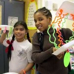
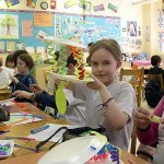
Catherine described how the students could build their own models of GPCRs, for hanging in the classroom as mobiles.

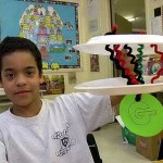
First, showing pictures of mobiles designed by their originator Alexander Calder, Catherine explained to the students to sculpt helices and loops from pipe-cleaners and thread them through two styrofoam trays that represent the inner and outer walls of the cell membrane.  Exploring protein folding in this fun way, the students placed finally a paper circle, with “G” written on one side and the student’s favorite odorous foods on the other side, onto the tail of their serpentine receptor models, to represent their associated G-protein.

With their GPCR models in hand, we reviewed how molecules bind to the loops outside of the cell, how such binding causes the helices to move inside the membrane and how such movement causes release of the G-protein to send to the inside of the cell a message, that something smells good outside.

For more information on GPCRs see:

http://en.wikipedia.org/wiki/G_protein-coupled_receptor


For signaling via G-protein-Coupled Receptors

www.web-books.com/MoBio/Free/Ch6D.htm


For an image of GPCRs and their importance as drug receptors see:

www.molres.org/jlameh/jlameh_gpcr.html


For more on Calder Mobiles see:

www.sfmoma.org/espace/calder/calder_windmobiles.html

www.guggenheimcollection.org/site/artist_work_md_26_2.html


Or our ‘Resource for Teachers’ poster : http://moleculesoflife.ca/2012/05/teach-about-g-protein-coupled-receptors-gpcrs/